
The First Signs of Organ Formation and the Blueprint of the Human Body
A New Chapter Begins
By the fifth day after fertilisation, the microscopic human embryo has reached one of the most remarkable milestones in early human development. Although still smaller than the tip of a pin, it has undergone an extraordinary transformation from a single fertilised cell into a highly organised collection of specialised cells. Every division that has taken place since fertilisation has followed an intricate genetic program encoded within the DNA.
This stage marks the appearance of the blastocyst, a sophisticated biological structure uniquely designed for survival, implantation, and future development. The embryo is now preparing for one of the most critical events of pregnancy—the successful attachment to the mother’s uterus. Without this event, development cannot continue.
The fifth day is therefore not simply another point in embryonic growth. It is the moment when the embryo begins to establish the foundation for every organ, tissue, and body system that will eventually form the complete human being.
From Morula to Blastocyst – A Remarkable Transformation
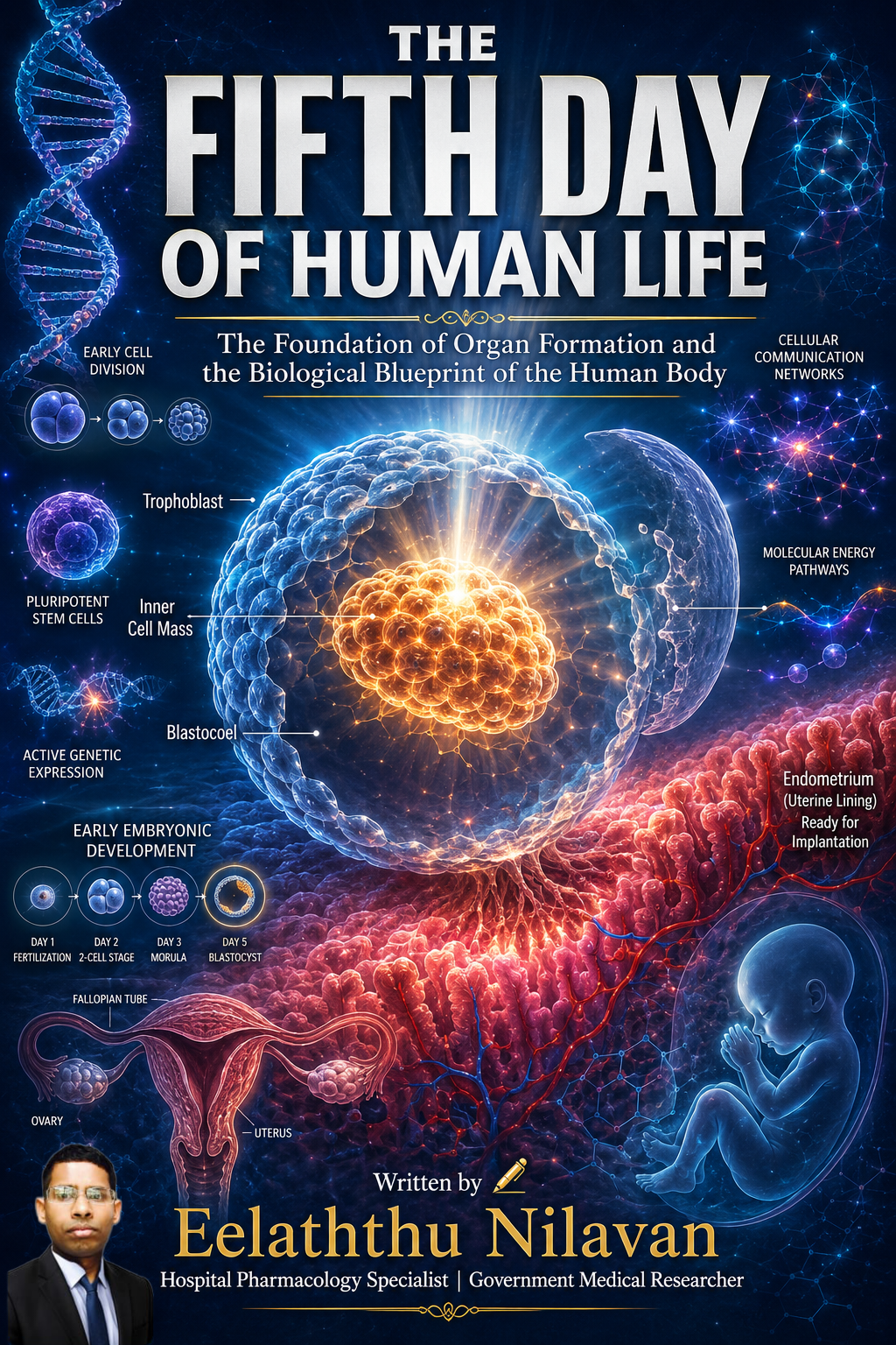
Over the previous four days, repeated cell divisions produced a compact ball of cells known as the morula. During the fifth day, these cells reorganise themselves into a hollow sphere filled with fluid. This new structure is called the blastocyst.
The blastocyst contains a fluid-filled cavity known as the blastocoel, which allows the cells to separate into distinct groups. This separation is essential because different cell populations are destined to perform completely different functions.
The embryo is no longer just multiplying cells. Instead, it is becoming a highly organised living system where each group of cells has a specific developmental destiny.
The Inner Cell Mass – The Beginning of Every Human Organ
One of the most important structures inside the blastocyst is the inner cell mass, also known as the embryoblast.
These remarkable cells will eventually develop into the entire human body, including:
• The brain
• The spinal cord
• The heart
• The lungs
• The liver
• The kidneys
• The digestive system
• Bones
• Muscles
• Skin
• Eyes
• Ears
• Every other organ and tissue
The cells within the inner cell mass are considered pluripotent stem cells, meaning they possess the extraordinary ability to transform into nearly every specialised cell type found in the human body.
Modern regenerative medicine continues to study these cells because of their enormous potential in treating neurological disorders, heart disease, diabetes, spinal cord injuries, and many degenerative conditions.
The Trophoblast – Building the Lifeline Between Mother and Baby
Surrounding the blastocyst is another important group of cells called the trophoblast.
Unlike the inner cell mass, trophoblast cells do not become part of the baby’s body. Instead, they develop into structures that support pregnancy, including the placenta.
The placenta serves as the baby’s life-support system throughout pregnancy by:
• Delivering oxygen
• Supplying nutrients
• Removing waste products
• Producing pregnancy hormones
• Protecting against many harmful substances
• Supporting immune tolerance between mother and fetus
Without a healthy trophoblast, a successful pregnancy cannot occur.
Hatching – Escaping the Protective Shell
Before implantation can begin, the blastocyst must escape from its outer protective covering, known as the zona pellucida.
This process is called hatching.
Special enzymes released by the embryo gradually weaken the zona pellucida until the blastocyst emerges completely free.
Hatching is essential because an embryo enclosed within the zona pellucida cannot attach to the uterine lining. Only after successful hatching can implantation proceed.
This represents one of the most delicate and crucial events in early embryonic development.
Preparing for Implantation
By the fifth day, the embryo begins searching for the optimal site within the uterus for implantation.
The lining of the uterus, called the endometrium, has been prepared by maternal hormones, particularly progesterone.
These hormonal changes transform the uterine lining into a soft, nutrient-rich environment capable of supporting embryonic growth.
Although implantation usually begins around days six or seven, the fifth day represents the final stage of preparation before this life-sustaining connection is established.
The Human Genome Takes Control
Another major developmental milestone has already occurred by this stage—the embryo’s own genome is now directing development.
Initially, embryonic growth depended largely on proteins and messenger RNA supplied by the mother’s egg.
By the fifth day, however, the embryo actively controls its own genetic program. Thousands of genes become activated, regulating:
• Cell communication
• Cell specialization
• Tissue organization
• Growth factors
• Metabolism
• Future organ formation
This marks the beginning of independent biological control over human development.
Cellular Communication – Every Cell Knows Its Role
The cells within the blastocyst constantly communicate using highly sophisticated molecular signals.
Chemical messengers regulate:
• Cell division
• Cell migration
• Gene activation
• Protein production
• Tissue organization
• Future body symmetry
These communication pathways ensure that each cell develops according to its precise biological function.
Even the smallest disruption in these signalling pathways may interfere with normal embryonic development.
Medical Importance of the Fifth Day
The fifth day has enormous importance in modern reproductive medicine.
During in vitro fertilisation (IVF), embryos are frequently cultured until they reach the blastocyst stage before being transferred into the uterus.
Blastocyst transfer offers several advantages:
• Improved implantation rates
• Better embryo selection
• Reduced risk of multiple pregnancies
• Higher success rates in many IVF programs
Embryologists carefully evaluate blastocysts based on their expansion, symmetry, and cell quality before selecting embryos for transfer.
Current Scientific Research
Scientists continue to investigate fifth-day embryos to better understand:
• Early pregnancy failure
• Infertility
• Recurrent miscarriage
• Genetic disorders
• Stem cell biology
• Placental diseases
• Congenital abnormalities
• Regenerative medicine
Advances in molecular biology, genetics, and developmental biology are expanding our understanding of these earliest stages of human life and may lead to new treatments for many diseases.
Conclusion – The Foundation of Every Human Life
The fifth day of human life is a profound moment in embryonic development. Within just five days of fertilisation, a single cell has become a highly organised blastocyst, containing the blueprint for every organ and tissue of the future human body. The embryo prepares for implantation, establishes distinct cell populations, activates its own genome, and lays the biological foundation for all subsequent growth.
Although invisible to the naked eye, the events of the fifth day demonstrate the extraordinary precision of human development. Every heartbeat, every breath, every thought, and every movement that will occur throughout life begins with these carefully orchestrated processes. The fifth day stands as a remarkable reminder that the complexity of human life is built step by step from its very earliest beginnings, guided by intricate cellular communication, genetic regulation, and the remarkable design of embryonic development.

Written by 𝐄𝐞𝐥𝐚𝐭𝐡𝐭𝐡𝐮 𝐍𝐢𝐥𝐚𝐯𝐚𝐧
Hospital Pharmacology Specialist | Government Medical Researcher